

X-Ray Technician
- Acquire the X-ray study
- Confirm study quality and views
- Release the study into XRAI
- Track intake status
- Resolve upload exceptions
Studies reach the worklist without repetitive manual uploads.
XRAI brings DICOM ingestion, AI-assisted analysis, primary care provider review and sign-off, digital signatures, final report generation, amendments, and workflow visibility into one connected radiology reporting experience.
XRAI supports the clinical workflow while qualified primary care providers remain responsible for reviewing, editing, approving, and signing reports.
XRAI in Action
Follow the complete workflow from study intake through AI-assisted analysis, provider review, signing, and final report delivery.
Workflow at a glance
01
A configured XRAI Agent detects and uploads supported DICOM studies.
02
Metadata registers the study in the shared worklist.
03
A provider claims, receives, or is assigned the study through the configured process.
04
Configured AI models return supporting observations, findings, or draft content.
05
The provider reviews images and AI output, then prepares the report.
06
An authorized provider reviews, approves, and signs where required.
07
The platform generates the final report and records its workflow state.
08
Authorized users add controlled amendments while preserving signed report history.
Unified Work Management
| Study Date | Patient | Study Description | Body Part | Claimed By | Report | Edit Report | Status |
|---|---|---|---|---|---|---|---|
15 Sep 2026 09:12 · XR-10842 | Sam Wahba 586320 · DOB 16/07/1974 | Lumbar spine and pelvis series | L SPINEPELVIS+1 | AM Dr. A. Morgan Idle · available | Awaiting My Signature | ||
15 Sep 2026 08:54 · XR-10841 | Marcus Reed 712904 · DOB 03/11/1968 | Chest PA and lateral | CHEST | NP Dr. N. Patel Idle · available | In review | ||
15 Sep 2026 08:37 · XR-10840 | Elena Torres 493812 · DOB 21/02/1986 | Left knee complete series | KNEE | JR Dr. J. Rivera Idle · available | Awaiting signature | ||
15 Sep 2026 08:05 · XR-10839 | Daniel Kim 834106 · DOB 09/09/1991 | Right wrist and hand | WRISTHAND | — | — | Analysis ready | |
14 Sep 2026 17:42 · XR-10838 | Priya Desai 621475 · DOB 12/05/1979 | Ankle and foot weight-bearing | ANKLEFOOT | EO Dr. E. Okafor Idle · available | Analysis ready · re-analysis | ||
14 Sep 2026 16:18 · XR-10837 | Olivia Chen 257963 · DOB 28/01/1958 | Pelvis and bilateral hips | PELVISHIP+2 | AM Dr. A. Morgan Idle · available | Amended | ||
14 Sep 2026 15:51 · XR-10836 | Noah Williams 908341 · DOB 07/06/2002 | Left foot complete series | FOOT | — | — | — | Analysis ready |
14 Sep 2026 15:14 · XR-10835 | Grace Miller 340178 · DOB 19/12/1965 | Chest single frontal view | CHEST | NP Dr. N. Patel Idle · available | — | Finalized | |
14 Sep 2026 14:33 · XR-10834 | Theo Bennett 175826 · DOB 24/08/1982 | Right knee | KNEE | JR Dr. J. Rivera Idle · available | Awaiting My Signature |
Scroll horizontally to view all workflow details
AI-Assisted Reporting
John Thompson
5019328
Diagnostic X-ray viewer
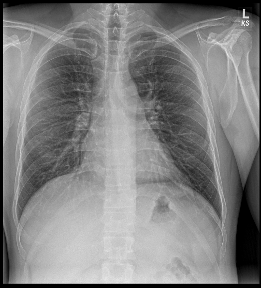
X-ray 1 of 2 · CHEST
Collaborative Reporting
Studies reach the worklist without repetitive manual uploads.
AI assists. The primary care provider interprets, signs and remains responsible for the report.
Operational teams maintain visibility without interrupting clinical review.
Before and After
See how XRAI can connect study intake, AI assistance, professional review, signing, report generation, and operational oversight in one governed experience.
Study in. Reviewed and signed report out.